top of page
Atrial Flutter: ECG Findings Explained
03:36
Medical Education for Visual Learners

Atrial Flutter: ECG Findings Explained

Atrial flutter is a macroreentrent dysrhythmia characterized by rapid flutter waves with a sawtooth appearance. The ectopic atrial depolarizations are called flutter waves and are usually best visualized in the inferior leads. The absence of an isoelectric line between the atrial flutter waves results in a ”sawtooth” appearance. The flutter rate is typically between around 250 and 350 per minute. Although the atrial rate can be very fast, typically only a portion of impulses are conducted to the ventricles. Physiologic conduction delay at the AV node usually limits the AV conduction ratio to between 2:1 and 4:1. If the AV conduction ratio is constant, then the rhythm will be regular. Otherwise the R-R intervals will be unequal. 📺 Subscribe To My Channel and Get More Great Quizzes and Tutorials https://www.youtube.com/channel/UC95TzSH1B_2EjaZMgDBNmvA?sub_confirmation=1 #FOAMed #cardiology #ECG Disclaimer: All the information provided by Medical Education for Visual Learners and associated videos are strictly for informational purposes only. It is not intended as a substitute for medical advice from your health care provider or physician. It should not be used to overrule the advice of a qualified healthcare provider, nor to provide advice for emergency medical treatment. If you think that you or someone that you know may be suffering from a medical condition, then please consult your physician or seek immediate medical attention.
Atrial Flutter: ECG Findings Explained
03:36
Medical Education for Visual Learners

Atrial Flutter: ECG Findings Explained

Atrial flutter is a macroreentrent dysrhythmia characterized by rapid flutter waves with a sawtooth appearance. The ectopic atrial depolarizations are called flutter waves and are usually best visualized in the inferior leads. The absence of an isoelectric line between the atrial flutter waves results in a ”sawtooth” appearance. The flutter rate is typically between around 250 and 350 per minute. Although the atrial rate can be very fast, typically only a portion of impulses are conducted to the ventricles. Physiologic conduction delay at the AV node usually limits the AV conduction ratio to between 2:1 and 4:1. If the AV conduction ratio is constant, then the rhythm will be regular. Otherwise the R-R intervals will be unequal. 📺 Subscribe To My Channel and Get More Great Quizzes and Tutorials https://www.youtube.com/channel/UC95TzSH1B_2EjaZMgDBNmvA?sub_confirmation=1 #FOAMed #cardiology #ECG Disclaimer: All the information provided by Medical Education for Visual Learners and associated videos are strictly for informational purposes only. It is not intended as a substitute for medical advice from your health care provider or physician. It should not be used to overrule the advice of a qualified healthcare provider, nor to provide advice for emergency medical treatment. If you think that you or someone that you know may be suffering from a medical condition, then please consult your physician or seek immediate medical attention.
Hypothermia ECG Explained
02:30
Medical Education for Visual Learners

Hypothermia ECG Explained

A classic ECG finding in hypothermia is J Point Elevation (a.k.a., Osborn J wave). Note the prominent convex deflection at the junction of the QRS complex and ST segment. It is most clearly evident in the precordial leads and typically negative in leads aVR and V1. The amplitude of the J wave roughly correlates with the severity of hypothermia. Although J waves are characteristic of hypothermia, they can also be present in Brugada syndrome, benign early repolarization, hypercalcemia, sepsis, and subarachnoid hemorrhage. Hypothermia can also cause diffuse slowing of impulse conduction. This can lead to prolonged intervals (e.g., PR, QT) and widened waveforms (e.g., p waves, QRS complexes). Inspect the QT interval in this ECG. The PR interval and P waves cannot be assessed due to the underlying rhythm. 
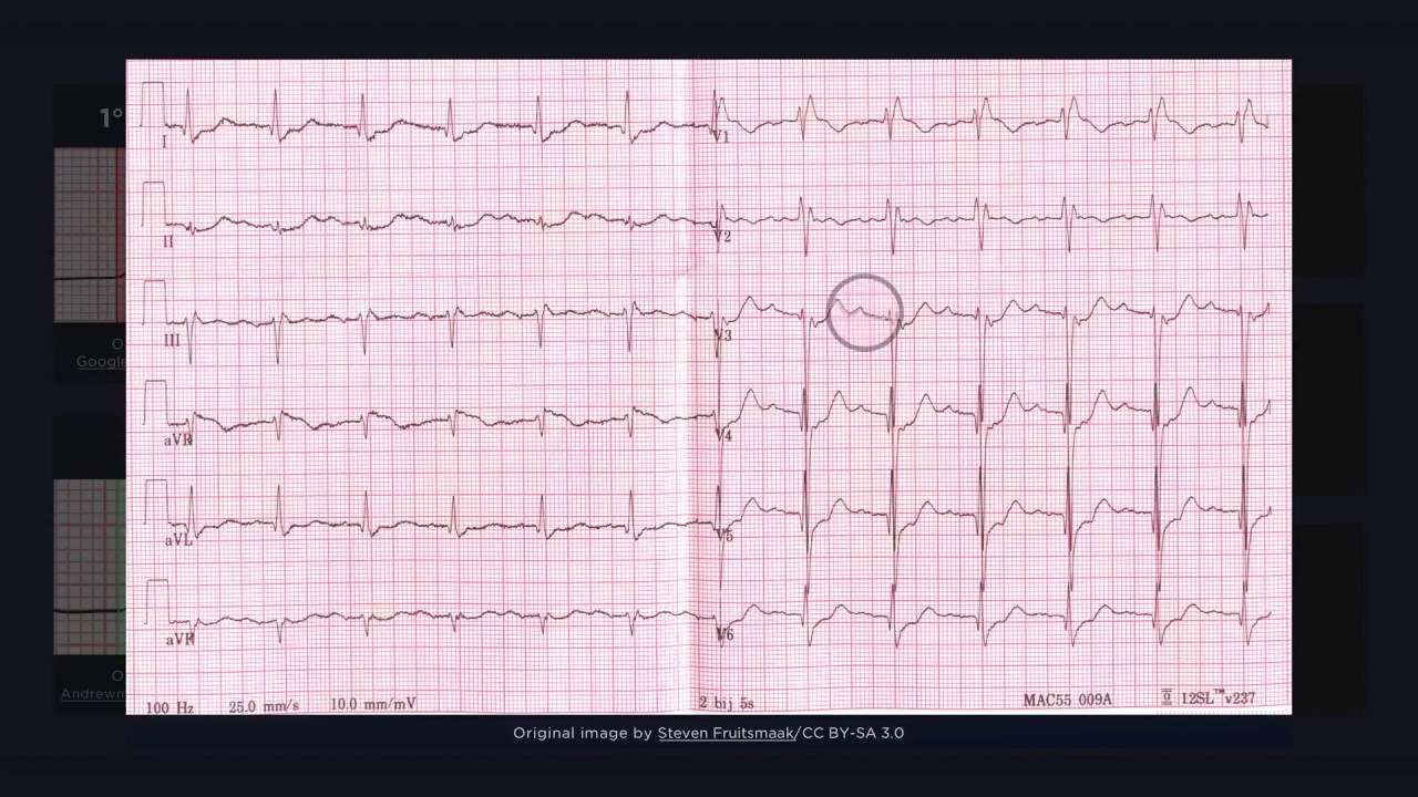
As well, marked slowing of the heart rate can occur due to a variety of bradyarrhythmias (e.g., sinus bradycardia, AV block, atrial fibrillation with slow ventricular response). In this ECG, Note the irregular R-R intervals and fibrillatory waves of A-Fib, yet the slow ventricular rate. One pitfall to be aware of, however, is that shivering can lead to electrocardiographic artefacts (e.g., waves simulating ventricular tachycardia). 0:00 J-point Elevation "Osborn J wave" 0:42 Slowed Conduction 1:08 Bradyarrhythmia 1:30 Pitfalls 📺 Subscribe To My Channel and Get More Great Quizzes and Tutorials https://www.youtube.com/channel/UC95TzSH1B_2EjaZMgDBNmvA?sub_confirmation=1 #FOAMed #cardiology #ECG Disclaimer: All the information provided by Medical Education for Visual Learners and associated videos are strictly for informational purposes only. It is not intended as a substitute for medical advice from your health care provider or physician. It should not be used to overrule the advice of a qualified healthcare provider, nor to provide advice for emergency medical treatment. If you think that you or someone that you know may be suffering from a medical condition, then please consult your physician or seek immediate medical attention.
Atrial Fibrillation ECG Explained
03:47
Medical Education for Visual Learners

Atrial Fibrillation ECG Explained

While looking at this ECG of atrial fibrillation, one of the first things that you might notice is that there aren’t any P waves. Instead, we can see a quivering baseline with interspersed QRS complexes. These atrial undulations are the result of rapid and erratic atrial depolarization and are referred to as fibrillatory waves. These fibrillatory waves can be either fine (i.e., less than 1 mm in amplitude) or coarse (i.e., greater than 1 mm in amplitude). When they are fine and have a low amplitude, they can appear as a smooth, wavy line or even a flat isoelectric line. These fibrillatory waves occur without any association to the irregularly dispersed QRS complexes. 0:00 Intro 0:22 Fibrillatory (f) waves 0:57 Irregular R-R Intervals 1:23 Calculating heart rate 2:17 No discernible p waves - Example 1 2:34 No discernible p waves - Example 2 2:48 Atrial fibrillation with left bundle branch block (LBBB) 3:11 Ashman phenomena Attribution: Original image of ECG rhythym in thumbnail by Drj / CC BY-SA 3.0. 📺 Subscribe To My Channel and Get More Great Quizzes and Tutorials https://www.youtube.com/channel/UC95TzSH1B_2EjaZMgDBNmvA?sub_confirmation=1 #FOAMed #cardiology #ECG Disclaimer: All the information provided by Medical Education for Visual Learners and associated videos are strictly for informational purposes only. It is not intended as a substitute for medical advice from your health care provider or physician. It should not be used to overrule the advice of a qualified healthcare provider, nor to provide advice for emergency medical treatment. If you think that you or someone that you know may be suffering from a medical condition, then please consult your physician or seek immediate medical attention.
Third Degree Atrioventricular Block ECG
03:00
Medical Education for Visual Learners

Third Degree Atrioventricular Block ECG

With third degree heart block, electrical impulses from the atria do not reach the ventricles. Instead, the atria and ventricles are controlled by independent pacemakers. This results in asynchronous activation of the upper and lower chambers of the heart. This is reflected on the ECG as atrioventricular dissociation, in which the P waves and QRS complexes occur completely independently of one another. The atrial rate is based on the underlying rhythm; in this case, the atria are under control of the sinus node. The P waves are simply not conducting to the ventricles, and thus do not initiate any QRS complexes. The QRS complexes that emerge are due to an escape rhythm, which is slower than the sinus rate. This is important, because AV dissociation by itself is not diagnostic of complete heart bock. For example, it could also occur in ventricular tachycardia. The morphology of the QRS complexes and the ventricular rate depends on the location of the escape pacemaker. When the block occurs at the level of the AV node and the escape pacemaker is in the AV junction, then a junctional escape rhythm appears, with a narrow (less or equal to 0.10 s) QRS complex and a rate usually between 40 and 60 beats per minute. When the block occurs below the level of the AV node and the escape pacemaker is in the ventricle, then a ventricular escape rhythm appears. The QRS complex is wide (more than 0.10 s) and the rate is usually about 20 to 40 beats per minute or less. Clinically, patients with ventricular escape rhythms are usually more compromised. 📺 Subscribe To My Channel and Get More Great Quizzes and Tutorials https://www.youtube.com/channel/UC95TzSH1B_2EjaZMgDBNmvA?sub_confirmation=1 #FOAMed #cardiology #ECG Disclaimer: All the information provided by Medical Education for Visual Learners and associated videos are strictly for informational purposes only. It is not intended as a substitute for medical advice from your health care provider or physician. It should not be used to overrule the advice of a qualified healthcare provider, nor to provide advice for emergency medical treatment. If you think that you or someone that you know may be suffering from a medical condition, then please consult your physician or seek immediate medical attention.
Atrioventricular Nodal Re-entry Tachycardia (AVNRT) ECG
03:39
Medical Education for Visual Learners

Atrioventricular Nodal Re-entry Tachycardia (AVNRT) ECG

AVNRT is a paroxysmal supraventricular tachycardia that occurs due to a functional re-entrant circuit within the nodal tissue. The electrical impulse goes round and round – typically down the slow pathway and up the fast - resulting in a regular rate of QRS complexes. The ventricular rate is rapid, usually between 140 and 280 beats per minute. Anterograde activation of the ventricles via the normal pathways from the AV node results in a narrow QRS complex. However, pre-existing aberrancy (e.g., left bundle branch block), can result in AVRNT rhythms with a wide QRS complex. Putting all that together, we could thus far describe our findings on this ECG as a fast, regular rhythm with narrow QRS complexes - all typical features of a supraventricular tachycardia. Now if you look closely, you will notice that there aren’t any p waves before the QRS complexes. Instead, there is a terminal deflection right after the QRS complex. 📺 Subscribe To My Channel and Get More Great Quizzes and Tutorials https://www.youtube.com/channel/UC95TzSH1B_2EjaZMgDBNmvA?sub_confirmation=1 #FOAMed #cardiology #ECG Disclaimer: All the information provided by Medical Education for Visual Learners and associated videos are strictly for informational purposes only. It is not intended as a substitute for medical advice from your health care provider or physician. It should not be used to overrule the advice of a qualified healthcare provider, nor to provide advice for emergency medical treatment. If you think that you or someone that you know may be suffering from a medical condition, then please consult your physician or seek immediate medical attention.
Second Degree AV Block ECG
03:52
Medical Education for Visual Learners

Second Degree AV Block ECG

Type one second degree AV block is characterized by progressive lengthening of successive PR intervals until an atrial impulse fails to conduct to the ventricles. The lack of conduction to the ventricles results in a pause in the rhythm and the absence of a QRS complex on the ECG. Notice how the baseline PR interval (i.e., the first PR interval following a non-conducted P wave) lengthens from beat to beat until a beat is dropped. Although it is obvious in this example, this finding can actually be quite subtle. In less conspicuous cases, it can help to compare the baseline PR interval -which is the shortest - with the PR interval preceding the non-conducted P wave – which is typically the largest. It is important to note that although baseline PR interval is usually normal in duration, it can be prolonged if there is a co-existing first degree heart block. Another finding typical of Mobitz I, which is hard to appreciate in this illustration, is an R-R interval that progressively shortens. The R-R interval is the longest following a dropped beat, and then shortens with successive beats until an atrial impulse fails to conduct to the ventricles. The ratio of atrial impulses that are conducted to the ventricles is usually between 3:2 and 7:6. However, this ratio can vary between cycles. Nonetheless, each group begins and ends with a P wave. In this example, the atrial-to-ventricular conduction ratio is 4:3. Type two second degree AV block is characterized by constant PR intervals and an intermittent failure of the atrial impulse to conduct to the ventricles. Unlike in Mobitz I, the PR intervals are consistent between all conducted beats. As well the R-R intervals are constant and the rhythm is regular until a beat is dropped. This can be referred to as a patterned irregularity. As in Mobitz I, the ratio of atrial impulses that are conducted to the ventricles is usually between 3:2 and 7:6. Again, this ratio can vary between cycles. With 2:1 AV block, there is only one PR interval within a grouping. The PR intervals between groups, however, are constant. As are the R-R intervals, unless the degree of block is variable. When the degree of AV block is greater than 2:1, the dysrhythmia is referred to as high grade – or advanced – AV block. High grade AV block can be confused with third degree AV block, however conducted P waves will have a consistent PR interval. 📺 Subscribe To My Channel and Get More Great Quizzes and Tutorials https://www.youtube.com/channel/UC95TzSH1B_2EjaZMgDBNmvA?sub_confirmation=1 #FOAMed #cardiology #ECG Disclaimer: All the information provided by Medical Education for Visual Learners and associated videos are strictly for informational purposes only. It is not intended as a substitute for medical advice from your health care provider or physician. It should not be used to overrule the advice of a qualified healthcare provider, nor to provide advice for emergency medical treatment. If you think that you or someone that you know may be suffering from a medical condition, then please consult your physician or seek immediate medical attention.
Torsades de Pointes: ECG Findings Explained
02:14
Medical Education for Visual Learners

Torsades de Pointes: ECG Findings Explained

With Torsades de Pointes, the QRS complexes are polymorphic with progressive changes in height, width, shape, and axis. A characteristic finding of torsades de pointes is a recurring reversal of waveform polarity. The gradual alteration in polarity gives the appearance of QRS complexes twisting around the baseline in a spindle-like fashion. Torsades de pointes is associated with QTc prolongation. Other common etiologies include electrolyte disturbances (e.g., hypokalemia, hypocalcemia, hypomagnesemia) and medication side effects (e.g., quinidine, amiodarone). The rapid and gradual alteration of QRS complexes can be mistaken for ventricular fibrillation. Ventricular fibrillation, however, is completely chaotic without any discernible pattern or similarity between adjacent waveforms. 0:00 Intro 0:11 Polymorphic Ventricular Tachycardia 0:19 Recurring reversal of waveform polarity 0:38 Example 1 1:01 Example 2 1:19 Recap Music: https://www.bensound.com 📺 Subscribe To My Channel and Get More Great Quizzes and Tutorials https://www.youtube.com/channel/UC95TzSH1B_2EjaZMgDBNmvA?sub_confirmation=1 #FOAMed #cardiology #ECG Disclaimer: All the information provided by Medical Education for Visual Learners and associated videos are strictly for informational purposes only. It is not intended as a substitute for medical advice from your health care provider or physician. It should not be used to overrule the advice of a qualified healthcare provider, nor to provide advice for emergency medical treatment. If you think that you or someone that you know may be suffering from a medical condition, then please consult your physician or seek immediate medical attention.
Wolff-Parkinson-White (WPW) Syndrome ECG
03:56
Medical Education for Visual Learners

Wolff-Parkinson-White (WPW) Syndrome ECG

Wolff-Parkinson-White Syndrome An accessory pathway bypasses the AV node and results in an early onset of ventricular depolarization (i.e. preexcitation). The lack of AV nodal delay is reflected on the ECG by a short PR interval that is less than 120 milliseconds in duration. Early depolarization of the ventricles occurs outside of the conduction system (i.e., via slow muscle fibre to muscle fibre conduction), which results in a slurred upstroke of the QRS complex. The QRS complex in WPW is wide because it is a composite of ventricular activation via both the bypass tract and the AV node. The initial portion of the QRS complex (i.e., the delta wave) is the result of aberrant ventricular depolarization via the accessory pathway, while the terminal portion reflects normal ventricular depolarization via AV node. A deep s wave in lead V1 indicates right to left-sided conduction; ventricular depolarization begins in the right side of the heart and then spreads towards the left ventricle. The ECG correlates of WPW syndrome may not be present during sinus rhythm and can also vary from day to day. As well, the WPW pattern can mask the findings that can occur with myocardial ischemia/infarction and other pathologies. 📺 Subscribe To My Channel and Get More Great Quizzes and Tutorials https://www.youtube.com/channel/UC95TzSH1B_2EjaZMgDBNmvA?sub_confirmation=1 0:00 Intro 00:11 Short PR Interval 00:41 Delta Wave 01:03 Wide QRS Complex 01:27 Right-Sided Bypass Tract (Type B pattern) 01:45 Pitfalls #FOAMed #cardiology #ECG Disclaimer: All the information provided by Medical Education for Visual Learners and associated videos are strictly for informational purposes only. It is not intended as a substitute for medical advice from your health care provider or physician. It should not be used to overrule the advice of a qualified healthcare provider, nor to provide advice for emergency medical treatment. If you think that you or someone that you know may be suffering from a medical condition, then please consult your physician or seek immediate medical attention.
bottom of page